Research

MDF 2019-2020 Fellow Shruti Choudhary Is Focused on Small Molecule Therapies for DM1

Published on Thu, 07/18/2019

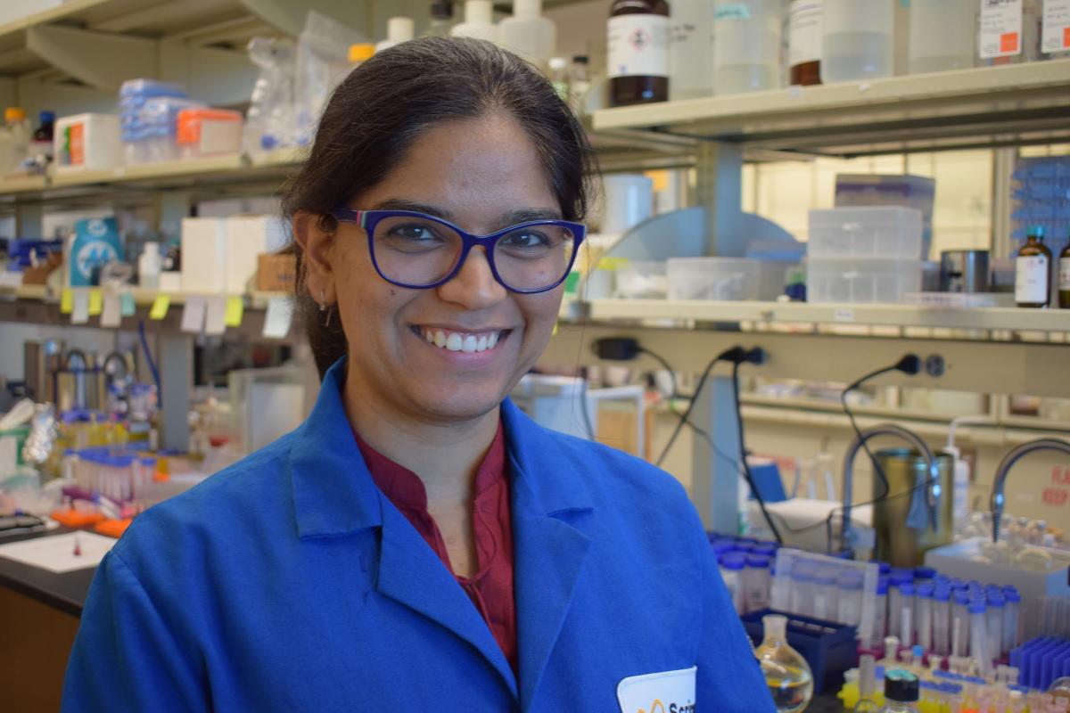
Shruti Choudhary, Ph.D., conducted doctoral work in medicinal chemistry at Duquesne University in Pittsburgh with a focus on cancer and infectious diseases, but once she had completed her degree, she decided to turn her attention to myotonic dystrophy. She felt there were plenty of disease targets, therapies, and researchers focused on cancer, and saw a far greater need to address neurodegenerative conditions. After hearing Dr. Matthew Disney from the Scripps Research Institute in Florida speak during a webinar, she applied to his lab for a postdoctoral fellowship, where she has worked since October 2017.

“My training was in chemistry and I wanted to get into a group that does both chemistry and biology,” she said. “There has to be an integration of both of these.”

MDF awarded Dr. Choudhary a 2019-2020 Fellowship to support her work in the Disney Lab exploring a potential therapy for DM1. The fellowship, awarded in conjunction with MDF UK, will provide two years of funding plus travel stipends to allow her to attend the annual MDF and biennial IDMC conferences.

The Disney lab has long worked on myotonic dystrophy and has identified certain small molecules that bind to RNA in a highly selective manner. They believe these molecules can be used to disrupt the disease process underlying DM1. Dr. Choudhary's work involves optimizing small molecule candidates to enhance their properties as potential therapies before putting them into in vitro (in a test tube, culture or otherwise outside a living organism) and in vivo (in a living organism) testing. She is modifying them to confer properties that would enhance their potential as drug candidates that could begin human clinical testing.

People with DM1 have what is known as a triplet repeat expansion in the non-coding portion of the DMPK gene. The gene codes for the production of myotonic dystrophy protein kinase, which is believed to play a critical role in muscle, heart, and brain cells. The expanded number of repeats in the DMPK gene results in the production of toxic RNA that also has an expanded number of repeats. Though it is normal for people to have a repeating piece of genetic code in both the DMPK gene and the resulting RNA, people with DM1 have an abnormally large and unstable number of repeats. Those repeats in the RNA bind to muscleblind-like proteins, master regulators of RNA, and prevent them from carrying out their normal functions.

The Disney Lab has hit upon a novel strategy to counter the ill-effects of the toxic RNA by appending small molecules that bind selectively to it, along with a nucleic acid-cleaving molecule part that cuts the disease-causing portion of the RNA and removes the toxic piece of code so proper cell activity can be restored.

“I am trying to see if I can make the structure smaller,” said Dr. Choudhary. “If the structure is smaller it will have better pharmacokinetic properties. That’s what I’m doing with this project right now.”

Small molecule drugs have traditionally been designed to bind to proteins, but Dr. Disney believes it is possible to develop small molecule compounds capable of binding to RNA, a novel strategy that provides new ways to approach a wide range of diseases.

Dr. Disney is a co-founder of Expansion Therapeutics, which was formed in 2016 to discover and develop small molecule drugs that target RNA. It secured $55.3 million in initial venture funding to conduct discovery and development of drugs focused on repeat expansion disorders with a lead program in DM1 and second one in discovery for DM2.

This is Dr. Choudhary's second attempt to become an MDF Fellow. Her first application was not selected, which made the notification that she had been selected this year much more exciting.

“It’s a huge honor and a huge responsibility,” she said.

Linking DM Molecular Events to Insulin Resistance and Muscle Atrophy

Published on Tue, 04/23/2019

Muscle metabolic defects have been suspected as contributors toward the pathogenesis of DM. Recent studies have suggested that cellular/molecular mechanisms underlying reduced insulin sensitivity in skeletal muscle of both DM1 and DM2 may be more complex that has been appreciated. Specifically, prior findings suggested that perturbations of post-INSR signaling may be a key factor in development of insulin resistance (Renna et al., 2017). A new publication builds on these data to better establish the link between DM splicopathy and the development of insulin resistance and skeletal muscle atrophy.

Linking DM Molecular Biology to Metabolic Dysfunction

Drs. Laura Renna, Giovanni Meola, and Rosanna Cardani and colleagues (IRCCS-Policlinico San Donato and University of Milan) investigated events downstream from DM-associated mis-splicing of the insulin receptor (INSR) to identify pathways underlying insulin resistance and skeletal muscle wasting (Renna et al., 2019). Specifically, the team asked whether a lack of insulin pathway activation could contribute to skeletal muscle atrophy. Dr Renna was the recipient of an MDF Fellowship in support of this work.

The research team evaluated muscle biopsies from 8 DM1 and 3 DM2 genetically confirmed patients enrolled in a national registry. Biopsies were assessed for muscle fiber type and morphometrics, insulin receptor protein expression, INSR alternative splicing, and signaling pathway response to insulin stimulation.

Skeletal muscle atrophy was detected in nearly all DM1 (type 1 and/or type 2 fiber atrophy) and DM2 (type 2 fiber atrophy) patients. In contrast to the pattern seen in controls (including samples from unaffected controls, motoneuron disease, and type 2 diabetics), RT-PCR analysis showed predominance of the fetal insulin receptor isoform transcript in both types of DM. When examined by fiber type, DM skeletal muscle exhibited a lower expression of insulin receptor protein than all controls for type 1 (slow oxidative) muscle fibers. The team found a negative correlation between type 1 fiber insulin receptor protein level and level of fetal insulin receptor transcript.

A means of assessing insulin pathway activation was identified and validated by the research team. Their results showed that defective activation of insulin signaling led to lower activation of mTOR accompanied by increases in MuRF1 and Atrogin-1/MAFbx expression—all key regulators known to act through insulin signaling to modulate skeletal muscle mass.

Modeling the Impact on Skeletal Muscle in DM

Taken together, these findings further advance understanding of the linkage between insulin receptor mis-splicing, metabolic dysfunction, and skeletal muscle atrophy. In terms of the insulin receptor, predominance of the fetal insulin receptor isoform in DM was observed, and an overall reduction in insulin receptor protein level in DM skeletal muscles was linked to reductions in receptor expression in type 1 fibers.

Based on these data, the research team suggests that reduced insulin receptor levels are responsible for the observed defect in insulin pathway activation in DM and a metabolic imbalance in protein synthesis/degradation—these, in turn, indicate a potential link to skeletal muscle weakness and atrophy, but causality has not yet been established.

Finally, another recent publication (Vujnic et al., 2018) expands upon this theme of metabolic dysfunction in DM, reporting an increased incidence of metabolic syndrome (i.e., co-occurrence of at least 3 of 5 symptoms: central obesity, high blood pressure, high blood sugar, high serum triglycerides, and low serum high-density lipoprotein) in DM2. Thus, there’s a clear case for increased research and patient management efforts directed toward metabolic dysfunction in DM.

References:

Receptor and post-receptor abnormalities contribute to insulin resistance in myotonic dystrophy type 1 and type 2 skeletal muscle.
Renna LV, Bosè F, Iachettini S, Fossati B, Saraceno L, Milani V, Colombo R, Meola G, Cardani R.
PLoS One. 2017 Sep 15;12(9):e0184987. doi: 10.1371/journal.pone.0184987. eCollection 2017.

Aberrant insulin receptor expression is associated with insulin resistance and skeletal muscle atrophy in myotonic dystrophies.
Renna LV, Bosè F, Brigonzi E, Fossati B, Meola G, Cardani R.
PLoS One. 2019 Mar 22;14(3):e0214254. doi: 10.1371/journal.pone.0214254. eCollection 2019.

Metabolic impairments in patients with myotonic dystrophy type 2.
Vujnic M, Peric S, Calic Z, Benovic N, Nisic T, Pesovic J, Savic-Pavicevic D, Rakocevic-Stojanovic V.
Acta Myol. 2018 Dec 1;37(4):252-256. eCollection 2018 Dec.

Targeting Toxic RNA in DM1 with a Small Molecule Drug

Published on Tue, 04/23/2019

As a proximate mediator of the splicopathy that characterizes DM1, DMPK RNA carrying the toxic expanded CUG repeat sequence represents a potentially important target for therapy development. A candidate therapeutic with drug-like properties, including the ability to access the many affected target tissues in DM1, that specifically targets expanded repeat RNA, would represent an important compound for evaluation in interventional clinical trials. A new study has taken steps to establish preclinical proof of concept for such an agent.

Initial Development of Cugamycin

Ms. Alicia Angelbello (PhD graduate student) and Dr. Matt Disney at The Scripps Research Institute, Florida and their colleagues (at University of Florida and Iowa State) have developed a small molecule compound capable of cleaving DMPK expanded repeat RNA, as evidenced by data from studies in DM1 patient-derived myotubes and a DM1 mouse model (Angelbello et al., 2019). The candidate therapeutic has been named Cugamycin, because of its specificity/affinity for expanded, but not sub-disease threshold, CUG repeats.

The research team explored the idea that bringing specificity to a known RNA cleaving compound, bleomycin A5, represents a rationale path toward a DM1 therapeutic. By attaching bleomycin A5 to an RNA-binding molecule, the team could achieve a critical level of specificity and thereby advance both bioactivity/efficacy and safety of the resulting compound—Cugamycin. Target recognition and cleavage specificity of Cugamycin was established through studies showing in vitro binding and efficient cleavage of CUGexp, but not DNA repeat hairpins.

Cell- and Animal-Based Support for Further Development of Cugamycin

In vitro analyses using DM1 patient-derived and control myotubes showed that Cugamycin was cell-permeable (overcoming a hurdle seen in large molecule development for DM1 muscle) and tracks to the nucleus. DMPK RNA cleavage efficiency in this model was 40%, with an EC25 in high nanomolar range—notably, wild type DMPK RNA was spared. The research team also established that Cugamycin reduced nuclear foci and rescued defects in the splicing events tested in the cell model. Direct comparison of Cugamycin and antisense oligonucleotides (AONs) targeted to DMPK expanded repeat RNA showed higher specificity for the small molecule drug (AON sequence/chemistry used here failed to distinguish between expanded and subthreshold DMPK RNA).

Findings of subsequent in vivo evaluations of drug metabolism and pharmacokinetics (the other DMPK) of Cugamycin supported a move to mouse efficacy testing. Short-term dosing of 10 mg/kg ip every other day in adult HSALR mice was well tolerated and without side effects linked to the base bleomycin A5 compound. Short-term dosing also produced a 40% reduction in toxic DMPK RNA and reversal of splicing defects tested in hindlimb muscles, confirming systemic drug bioavailability and target engagement and modulation. Treated HSALR mice showed restoration of Clcn1 protein levels and reductions in myotonia. The high selectivity of the candidate therapeutic was confirmed in RNA-seq analyses of skeletal muscle samples from untreated wild type and vehicle only and Cugamycin treated HSALR mice.

The authors conclude that, as a preclinical candidate molecule, Cugamycin has limited liabilities, but do note the opportunities for further chemical analoging around its scaffold to continue to optimize and arrive at a development candidate for IND-enabling studies and entry into clinical trials.

Path Forward

Taken together, this new study provides a compelling case that small molecules can be developed to safely and effectively target toxic RNA cleavage in DM1. At the time of this writing, yet another reminder became available that efficacy in a mouse model may or may not translate into an effective drug in patients (Chakradhar, 2019 and justsaysinmice). Thus, while current findings are encouraging, they are in a model organism that cannot actually “have” the human disease—we look forward to further testing of Cugamycin and its analogs, and pursuit of all strategies for the development and regulatory approval of safe and effective drugs for patients living with DM.

References:

Precise small-molecule cleavage of an r(CUG) repeat expansion in a myotonic dystrophy mouse model.
Angelbello AJ, Rzuczek SG, Mckee KK, Chen JL, Olafson H, Cameron MD, Moss WN, Wang ET, Disney MD.
Proc Natl Acad Sci U S A. 2019 Mar 29. pii: 201901484. doi: 10.1073/pnas.1901484116. [Epub ahead of print]

It’s just in mice! This scientist is calling out hype in science reporting.
Chakradhar, S.
STAT. April 15, 2019
 

Potential Involvement of RNAi Mechanisms in DM1

Published on Tue, 04/23/2019

The muscular dystrophy field provides case studies of how flawed assumptions about the mechanistic understanding of a disease can lead therapeutic discovery and development efforts astray. One should never assume that we “already know” all key aspects of disease pathogenesis and thus have identified all feasible targets for therapeutics.

While the DM field certainly has developed a strong disease understanding, including of molecular targets that are tractable for drug and biologic development, there are many nagging questions remaining, such as the role of RAN translation in DM1 and DM2 and that of the epigenetic modifications upstream of the DMPK locus in CDM. In addition to these known unknowns, there likely are unknown unknowns in DM biology to worry about. Therapeutic efforts certainly need to go forward for rationale targets. But, as the NINDS concluded several years ago, there will always be a need for the novel findings that originate only from continuing basic and mechanistic research.

Might RNAi Machinery Play a Role in DM1?

A new article (Qawasmi et al., 2019) from Drs. Susana Garcia (University of Helsinki), Yuval Tabach (The Hebrew University of Jerusalem), and colleagues explores an alternative gain-of-function mechanism, that expanded CUG repeats in the DMPK transcript serve as templates for gene silencing via RNA interference (RNAi). This work utilized a novel C. elegans model of DM1.

The research team used the worm model they had developed previously, expressing 123 CUG repeats in the 3’UTR of a GFP transcript driven by a skeletal muscle promoter. Phenotypically, this model is characterized by motility and heat shock survival response defects.

To show that RNAi could be activated by expanded CUG repeats independent of other RNA mechanisms known to be operative in DM1, wild type worms were fed plasmid expressing RNA with 50 CTG repeats—this RNA is known to be cleaved and the resulting short CUG fragments activate RNAi. Data showed the development of motility defects similar to that of their 123 CUG DM1 model and a heat shock survival defect.

Consistent with findings in wild type worms fed expanded CUG plasmid, the 123 CUG repeat DM1 model showed, over time, a decay of the exogenous CUG repeat transcript and its protein product. By silencing RNAi pathway genes, the team showed that gene silencing was responsible for disappearance of the 123 CUG repeat. In the next series of experiments, they tested whether CUG repeats in their DM1 model were similarly processed and act as non-coding RNAs to silence expression of other endogenous CUG-bearing genes via RNAi. In these studies, expression of endogenous genes bearing at least 4 repeats was reduced by 1.6-fold. This suggests that expanded CUG repeat transcripts activate RNA silencing followed by the downregulation of endogenous CUG-bearing transcripts. Using RNA-seq of entire worms, a total of 982 genes were found to be downregulated by ≥ 1.5x; because some of the knocked down genes are not expressed in muscle, the overall effect apparently involves non-cell-autonomous RNA silencing in adjacent tissues. Finally, knocking down genes comprising the RNAi machinery rescued expression of genes that were otherwise affected in the 123 CUG repeat model.

Connecting the Dots

Collectively, these studies support the conclusion that expression of CUG expanded repeats triggers an RNAi-mediated repression of multiple endogenous genes that normally contain CUG repeats in their transcript. Thus, RNAi pathways may contribute towards the pathogenesis of DM1 and thus may represent a potential target for therapeutic intervention in the disease. Future studies should evaluate the potential operation of RNAi driven pathology in mammalian models of DM1 and determine its relative contribution to disease.

Reference:

Expanded CUG Repeats Trigger Disease Phenotype and Expression Changes through the RNAi Machinery in C. elegans.
Qawasmi L, Braun M, Guberman I, Cohen E, Naddaf L, Mellul A, Matilainen O, Roitenberg N, Share D, Stupp D, Chahine H, Cohen E, Garcia SMDA, Tabach Y.
J Mol Biol. 2019 Mar 14. pii: S0022-2836(19)30121-4. doi: 10.1016/j.jmb.2019.03.003. [Epub ahead of print]

CNS and Myotonic Dystrophy: Outcome Measures Development Workshop

Published on Tue, 04/23/2019

MDF will host a Workshop on CNS involvement in DM and the development of outcome measures for CNS-targeting therapies on September 12, 2019, in conjunction with the MDF Annual Conference, September 13-14, in Philadelphia, PA.

This Workshop will examine the natural history of DM1, DM2 and CDM and the underlying scientific premise for targeting therapy development to address the CNS phenotype, a key contributor to overall burden of disease. Through platform presentations and focused discussion sessions, the Workshop will seek to identify targetable CNS phenotypes, evaluate the current knowledge of the underlying disease mechanisms and molecular targets, and discuss what biomarkers and outcome measures might best demonstrate the clinical effectiveness of drugs and biologics in patients living with DM1, DM2 and CDM.

The meeting will take place from 8:00 AM to 4:00 PM. Lunch will be provided.

Click here to see the working agenda.

Registration Open Now

The Workshop agenda is currently under development. Interested parties are encouraged to register for the CNS Workshop and the 2019 MDF Annual Conference.

Annual Nationwide Children’s Hospital/OSU Myology Course

Published on Tue, 04/23/2019

Nationwide Children’s Hospital and Ohio State University have operated a 5-day myology training course for the last seven years. The course includes common lectures in the mornings and separate clinical and laboratory tracks in the afternoons. MDF staff have participated as course lecturers in the past and attest to the value of the course. Trainees accepted for the course pay only for travel and some meals, as there is no cost for the course itself or for local lodging. Attendance is capped at 60 (30 in each track), so those interested need to register quickly.

A slightly shortened version of the course announcement from Dr. Kevin Flanigan is provided here.

“I would like to invite you to send your trainees to the eighth annual Nationwide Children’s Hospital/Ohio State University Myology Course, to be held at Nationwide Children's Hospital from Monday, August 26th through Friday, August 30, 2019. The goal of this course is to provide trainees with an up-to-date and expert survey of neuromuscular diseases. The expert faculty will address clinical syndromes, mechanisms of pathogenesis, molecular therapeutics, and aspects of career development.

“The course is available to both pre-and post-clinical trainees, and both clinical- and laboratory-based trainees are welcome. We have structured the course so that both groups will be together for morning lectures.

“The Clinical Track is primarily directed at clinical fellows (in Neuromuscular Disease; Genetics; Electrophysiology; Physical Medicine & Rehabilitation; etc.), although other specialties and levels of training will be considered.

“Trainees in the Laboratory Track will have electives to choose from for the afternoon lab courses.

“The course is sponsored by Parent Project Muscular Dystrophy and the Muscular Dystrophy Association.

Register at http://bit.ly/19MYO. In order to register for this course, the trainee must (1) fill out the form at the link, and (2) submit a CV or biosketch to kevin.flanigan@nationwidechildrens.org. I will confirm their eligibility for the course. Their registration will not be complete until they receive confirmation from me of their acceptance.”

Pharma/Biotech Updates

Published on Tue, 04/23/2019

Recent announcements from three biotechnology and pharmaceutical companies reflect the increasing interest in and tractability of myotonic dystrophy for therapy development. Short summaries and links to the press releases are provided here.

AMO Therapeutics

AMO Therapeutics is in clinical testing of AMO-02 for treatment of congenital and childhood onset myotonic dystrophy. Their strategy is to target GSK-3β with a small molecule drug in order to address the considerable CNS burden of disease.

To date, the large majority of therapeutic development efforts in myotonic dystrophy have targeted skeletal muscle. The ease of accessing and evaluating splicing events in small muscle biopsies provides a drug developer with rather rapid insights as to drug bioavailability and molecular target modulation. Assessment of the CNS in myotonic dystrophy patients is more complex—the hurdles here include identifying appropriate drug targets and targetable CNS phenotypes and determining how interventional trials can best demonstrate the clinical effectiveness of drugs and biologics in patients. Because of the importance of the CNS phenotype in DM, MDF is devoting a workshop to this topic, scheduled for September 12, 2019.

In efforts to establish sensitive and effective CNS outcome measures for congenital myotonic dystrophy, AMO Therapeutics has developed the Clinician-Completed Congenital Myotonic Dystrophy Type 1 Rating Scale (CDM1-RS). The new scale builds upon prior efforts on clinical rating scales by DM researchers, Drs. Chad Heatwole and Nicholas Johnson. CDM1-RS is currently being validated in a natural history study in children and adolescents with DM1 and AMO plans using it as the primary outcome measure in their upcoming registration trial.

The full press release is available on the AMO website.

Audentes Therapeutics

Audentes Therapeutics has announced the licensing of AT466 from Nationwide Children’s Hospital (Drs. Kevin Flanigan and Nicolas Wein) to target DM1 via facilitated delivery of an oligonucleotide therapeutic. Efforts are currently in the preclinical stage; the company notes that IND filing is planned for 2020.

The development of large molecule drugs, such as oligonucleotides, has a critical barrier to overcome in muscle diseases—the rather poor uptake of these large molecules by intact skeletal muscle fibers. Under evaluation is a strategy to deliver and enhance oligonucleotide uptake via AAV vectors. Two categories of oligo will be tested to block accumulation of toxic expanded repeat DMPK RNA in cells—oligos to achieve RNA knockdown and others for exon skipping—to treat DM1.

The full press release is available on the Audentes website.

Dyne Therapeutics

Earlier this month, Dyne Therapeutics announced that they had raised $50M in Series A financing in support of their lead disease target, DM1. Funding will support advancing a development candidate into clinical testing—a putative timeline has not yet been provided.

The development of large molecule drugs, such as oligonucleotides, has a critical barrier to overcome in muscle diseases—the rather poor uptake of these large molecules by skeletal muscle fibers. Dyne proposes to address this hurdle by linking oligonucleotides to an antibody that tracks to and is internalized via muscle cell surface receptor binding. Once this facilitated uptake process gains entry of the oligonucleotide into muscle fibers, the Dyne oligonucleotide mechanism is degradation of toxic expanded CUG repeat RNA.

The full press release is available on the Dyne Therapeutics website.

Dr. Vincent Dion Awarded a $250,000 MDF Grant to Pursue Cure for DM

Published on Thu, 04/11/2019

First Grant in $1M DM Cure Development Project Awarded

MDF community members likely remember that in late 2017 very generous donors committed $1M to MDF to launch a gene editing development project to find a cure for myotonic dystrophy. MDF conducted a comprehensive scoping and discovery workshop, followed by a Request for Applications in mid-2018. We are delighted to announce that Dr. Vincent Dion was awarded the first grant for that project.

Dr. Dion's Approach

Dr. Dion was trying to understand why the size of the repeating piece of genetic code in the gene that causes myotonic dystrophy varied from cell to cell in patients with the condition and in so doing, he may have stumbled on a novel treatment for the disease.

MDF has awarded Dion a two-year grant totaling $250,000 to support his research to determine the feasibility of a gene editing treatment for DM. He will be helped by Drs. Geneviève Gourdon at the Imagine Institute in Paris, and Jack Puymirat at Université Laval.

Myotonic dystrophy is known as a trinucleotide repeat disorder because of an expanded repeat of a three-letter piece of genetic code CTG (cytosine, thymine, and guanine) found in the DMPK gene of people with DM1. It’s normal to have several repeats of this code in the DMPK gene, but people who have more than 50 repeats, can develop DM1. The greater the number of repeats, the more severe the disease tends to be.

A Surprising Finding

Dion, a professor at the UK Dementia Research Institute at Cardiff University, knew that the size of repeats in any person with the disease could vary greatly from cell to cell. He wanted to understand why. He was using the gene editing technology CRISPR-Cas9, a bacterial enzyme that allows users to make precisely-targeted cuts in genetic material, to see if he could test a theory that changes in the number of repeats was caused by DNA damage. He wanted to see if disrupting the repeat track by making cuts in a single track of the double-stranded genetic code to expose the repeat would influence whether expansion of the repeat occurred.

In a surprising finding, Dion observed that by making such breaks in the repeat track he had corrected the error. Enzymes that naturally repair damaged DNA healed the cuts to the repeat track. But instead of amplifying the repeat, it reduced its size.

In essence, his work suggested gene editing might prove to be a way to correct the toxic repeat and possibly arrest the mechanism of the disease.

“We stumbled upon this by chance,” said Dion, who earned his Ph.D. in molecular biology at Baylor College of Medicine in Houston and then completed a postdoctoral fellowship at the Friedrich Miescher Institute in Basel. “The question we were asking was completely different and we ended up using CRISPR to correct the mutation in human cells, and then we went from there.”

Accelerating Research with MDF

The MDF grant will allow Dion to continue his work, exploring the gene editing approach in a DM mouse model to see if it corrects the mutation.

The grant will also be used to see how precise the approach will be within a living organism and to make sure that the CRISPR cuts the expanded repeat in the DMPK gene as intended, and not elsewhere, which could lead to severe off-target effects.

Dion does have some tricks to ensure the CRISPR targets the genetic material correctly at the desired point. He uses RNA to target the repeat track he wants it to cut. That’s not without its challenges, as he needs to package the CRISPR and RNA into a viral vector to carry it into the cell.

The other issue any gene editing therapy will face is the need to deliver it into the central nervous system (the brain) to address the neurological effects of the condition. That would likely require some means of direct delivery. In the mouse model, Dion will be injecting directly into the brain. If all of that works, Dion then hopes to move the gene editing work to a larger DM animal model.

For now, Dion hopes to demonstrate that the approach works. Even if he is successful with the experiments he'll be conducting over the next two years, he believes it would still be several years before the approach could advance to human testing.

“The hope is that we'll know whether it works. We'll know whether it's specific enough for the repeat track and whether it might cause other mutations,” said Dion. “If those questions are answered successfully, then I think we're looking at another five years or so after that to do a clinical trial.”

Sleep and Disordered Breathing in DM2

Published on Tue, 03/19/2019

Although excessive daytime sleepiness and sleep disordered breathing are well documented for patients living with DM1, there is less clarity regarding these symptoms in DM2. A new review article in Current Neurology and Neuroscience Reports examines the literature on sleep and breathing disorders in DM2. Respiratory muscle weakness, myotonia, and alterations in central nervous system respiratory centers are believed to be causative.

Current Status of DM2 Research

One of the most significant gaps in understanding of myotonic dystrophy is the relative dearth of natural history studies in DM2. While there is not yet an approved, disease-mitigating therapy for myotonic dystrophy, clinical trial readiness has vastly improved for DM1, but not for DM2. It is critically important that DM2 cohorts be recruited and phenotyped in order to understand the longitudinal progression of the disease and thereby identify biomarkers and clinical assessment measures that will enable clinical trials in this patient population. If phenotypic data do not become available and DM2 is not perceived as ‘tractable’ by the pharma and biotech industry communities, it will be difficult to convince companies to address this disorder.

Sleep Complaints and Breathing Disorders in DM2

Dr. Andrea Romigi (IRCCS Neuromed, Italy) and colleagues recently published a review article looking at literature focused on breathing and sleep disorders in patients with DM2 (Romigi et al., 2019). Sleep disorders are important contributors to the morbidity and mortality of this disease and these findings are important in establishing clinical trial readiness for DM2.

Both skeletal muscle weakness and CNS dysfunction are believed to contribute to respiratory dysfunction characteristic of DM2, but careful assessments of lung function in DM2 have not been common. The most commonly reported sleep disorders in DM2 are sleep-disordered breathing (38–67%) and restless legs syndrome (50–60%). Sleep disordered breathing, in turn, appears to be the major cause of excessive daytime sleepiness in DM2. Claims as to the value of non-invasive ventilation appear to have an almost anecdotal basis and have not yet been sufficiently studied in DM2.

Restless legs syndrome has been viewed as significant in DM2, although this finding appears to have its basis only in studies of small cohorts. The authors of this review suggest that the prevalence of restless legs syndrome may be overstated by the frequent occurrence of myalgia and pain in this patient population, although they also note that the PRISM-2 study reported fatigue in 89% and excessive daytime sleepiness in 77% of DM2 patients.

Taken together, the authors of this review advocate for the inclusion of sleep studies in evaluation of patients with DM2. While data suggest that the frequency of hypoventilation and pulmonary restriction in DM2 is low, they also suggest that use of standard pulmonary examinations may improve upon disease morbidity. Finally, the potential correlation of pain, that is so characteristic of DM2, and sleep disruption has not been fully studied and should be an important component of future natural history studies of patients with DM2.

Reference:

Sleep Complaints, Sleep and Breathing Disorders in Myotonic Dystrophy Type 2.
Romigi A, Maestri M, Nicoletta C, Vitrani G, Caccamo M, Siciliano G, Bonanni E, Centonze D, Sanduzzi A.
Curr Neurol Neurosci Rep. 2019 Feb 9;19(2):9. doi: 10.1007/s11910-019-0924-0. Review.

Potential for Genome Editing in Neuromuscular Disease

Published on Tue, 03/19/2019

Over the last year, MDF has carefully examined the potential of genome editing strategies to address myotonic dystrophy, including hosting an expert workshop and a competition for research grant funding. In these efforts, the focus has been upon the need to evaluate various genome editing strategies, optimize reagents, identify an effective delivery vehicle, and establish systems to assess both safety and efficacy in preclinical models—as reported out from the workshop. Although genome editing is rapidly moving into the clinic for other disease indications, the technology likely is not yet optimized and the DM field will benefit from learnings from any first-in-man studies.

Progress in Genome Editing for Neuromuscular Diseases

Haris Babačić (now a Ph.D. student in proteogenomics at Karolinska Institute), Dr. Benedikt Schoser (Ludwig-Maximilians-University of Munich), and colleagues have conducted an intensive analysis of preclinical research publications in neuromuscular and repeat expansion disorders to ascertain key lessons for moving genome editing strategies forward. Their review was recently published in PLoS One (Babačić et al., 2019). Their review provides important perspectives obtained from one genome editing platform, CRISPR-Cas, so that learnings are not siloed within disease fields.

These authors utilized literature searches of MEDLINE and EMBASE databases (coverage up to the end of 2017) to identify publications focused on use of CRISPR-Cas genome editing systems in preclinical studies of at least one monogenic neuromuscular disease. In all, forty-two publications met all eligibility criteria and were reviewed. Like the MDF Genome Editing Workshop, this publication identified the efficient delivery of editing reagents and evaluation of their safety and efficacy as the salient issues in moving toward clinical testing of this therapeutic strategy.

The majority of studies reviewed here (n = 26) were in preclinical models of Duchenne muscular dystrophy (DMD); information was also obtained from publications in Huntington Disease (HD; 5 publications), Spinocerebellar Ataxia 2 (SCA2; 3 publications), Friedreich’s ataxia (FA; 1 publication), myotonic dystrophy type 1 (DM1; 3 publications), Fragile X syndrome (FXS; 2 publications), and three other types of muscular dystrophy (FSHD, MDC1A, and LGMD; 1 publication each).

Lessons from Studies of Genome Editing

The theme that the efficacy of genome editing is influenced by selection and delivery of guide RNAs (gRNA) and Cas was readily apparent among the publications focusing on DMD. Efficacy was shown for DMD in in vitro, ex vivo, and in vivo models. Delivery predominantly relied upon viral vectors, although use of gold nanoparticles has shown potential. Evaluation of the specificity of off-target editing (safety) largely focused on sequences predicted using bioinformatic models—an approach unlikely to be sufficient to support clinical development (as per the NIST Genome Editing Consortium).

Studies in DM may need to overcome difficulties in removal of the long, expanded repeat tracts; yet a strategy of targeting and eliminating the toxic repeat RNA with dCas9 has also been tested. The review’s authors note that mRNA targeting would require life-long administration of the candidate therapeutic—potential consequences of that strategy are not yet known, although there is a more general concern about off-target and immunological consequences of continues expression of the Cas protein. These safety concerns may require use of a delivery system in which Cas protein can be switched off after editing is complete.

Analysis of the other indications addressed in this review led to few additional lessons, outside of therapeutic window considerations that are disease-specific. The authors do include an important section on ethical issues that accompany genome wide editing strategies, ranging from benefit-risk assessments to the societal issues raised by germline editing. Overall, the review’s authors acknowledge the rapid progress that has been made in moving a potentially game-changing therapeutic forward, but conclude that the evidence to move into clinical trials for neuromuscular diseases is insufficient. They argue for more basic research to optimize delivery systems, thoroughly assess off-target safety concerns, and create standards for efficacy determination before transitioning to clinical trials. These are goals that are consistent with the due diligence that MDF has done to assess the opportunities and challenges of using genome editing in DM.

Reference:

CRISPR-cas gene-editing as plausible treatment of neuromuscular and nucleotide-repeat-expansion diseases: A systematic review.
Babačić H, Mehta A, Merkel O, Schoser B.
PLoS One. 2019 Feb 22;14(2):e0212198. doi: 10.1371/journal.pone.0212198. eCollection 2019.